Accelerators for Material characterisation
Accelerators for Material Characterisation
Material characterisation refers to a collection of techniques that are used to probe the internal structure of matter to gain a deeper understanding of its properties. Material characterisation is a key tool of materials science and has allowed for exciting developments in our understanding of materials. This has allowed researchers to develop new materials that could revolutionise the world we live in. These new advancements in materials science have potential applications in areas such as medicine, industry, alternative fuels and many others. Some potential techniques and applications of material characterisation are looked at in the sections below.
Cultural heritage, archaeology, dating and authentication
Techniques developed for fundamental research in materials science have found applications in many areas, including areas such as cultural heritage and archaeology. Examples include the use of Accelerator Mass Spectroscopy (AMS) to date archaeological finds or Ion Beam Analysis (IBA) to examine the composition of various layers of a painting.
Ion Beam Analysis
Ion Beam Analysis (IBA) is the name given to a large number of techniques, based on ion beams produced by particle accelerators to carry out material analysis. The beauty of IBA techniques is their ability to reveal the composition of any inorganic material without altering it, destroying it or removing samples from it all of which can be carried out in air (using accelerators often requires work to be carried out in vacuum). This makes IBA the ideal tool for the analysis of the inks, pigments, glass, ceramics, gems, stones or metal alloys of priceless works of art or archaeological finds. Techniques such as IBA, that do not damage samples, are collectively known as Non Destructive Testing (NDT). So powerful are these techniques that it is possible to reveal the palette that was used by an artist to create a work of art.
For more information on IBA see;
Web (University of Surrey): What is Ion Beam Analysis?, its applications
Doc [French]: Pleins feux sur les objets d'art et d'archéologie avec l'accélérateur de particules du Louvre, J.-C. Dran, Bulletin de la Société française de physique, 136, (2002)
See also: Article [2008] (CNRS): Vintage wine bottles authenticated by high energy ion beam
related video [French]: In Vitrum Veritas 8 min
Several facilities in the world use accelerators for ion beam analysis, for example AGLAE (Accélerateur Grand Louvre d'Analyse Elémentaire) located in C2RMF (Paris, France) and LABEC (Florence, Italy).
For more information see;
Web (eu-artech.org): Accélerateur Grand Louvre d'Analyse Elémentaire (AGLAE) on Eu-ARTECH (Access, Research and Technology for the conservation of the European Cultural Heritage)
Web: LABEC, Laboratorio di Tecniche Nucleari per i Beni Culturali – Firenze
Web (acceleratorer.com): Ion beam accelerators around the world
As mentioned previously, IBA covers a large number of complementary techniques. Rather than list all of these techniques, two examples of IBA techniques, Particle Induced X-ray Emission (PIXE) and Ion Beam Induced Luminescence (IBIL) are looked at below.
Particle Induced X-ray Emission (PIXE)
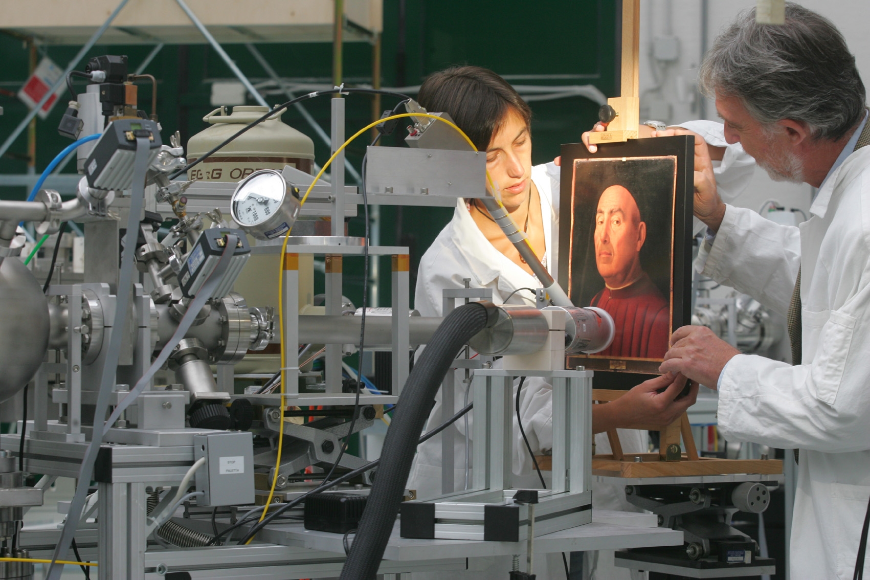
The most powerful IBA technique is PIXE, which relies upon the emission of characteristic X-rays from the investigated material, induced by ion beams. Two of the numerous advantages of the PIXE method are that it is non-destructive and allows for the quantitative examination of materials.
|
The “Ritratto Trivulzio” by Antonello da Messina during the analysis with particle accelerator. Image credit: LABEC, INFN's Laboratory for Cultural Heritage and Environment, Italy |
For more information on PIXE see;
Web (SPIRIT): Proton Induced Gamma-/X-ray Emission
Web (culture.gouv.fr) [French]: Analyse chimique par la méthode PIXE
The PIXE method is also used for studying atmospheric aerosols, which is addressed in the case study: Accelerators for a sustainable future.
This method finds also applications in forensic science. For example it enabled investigators to discover the truth of the ‘Markov Pellet’ spy story, where the Bulgarian dissident Georgi Markov was assassinated in London by a poison pellet umbrella. The pellet was later analysed and found to contain the poison ricin.
For more information see the Lecture: Ion Beam Analysis for Forensic Science referring to the document Farewell to Harwell’s Hangers (UKAEA)
Ion Beam Induced Luminescence (IBIL) / Iono-luminescence (IL)
Iono-luminescence (IL) or Ion Beam Induced Luminescence (IBIL) is an IBA technique in which a material is irradiated by an ion beam and made to emit light. IL is able to provide information about the chemical form of elements, which cannot be obtained by other ion beam analytical methods.
For more information on IBIL see;
Web (Leipzig University): The Ionoluminescence (IL) method
Using Synchrotron Light
Synchrotron light sources are particle accelerators built to accelerate charged particles in curved paths so as to produce high energy electromagnetic radiation known as Synchrotron radiation. Synchrotron radiation is used for numerous applications and in particular to perform analysis on various important cultural and historical artefacts. These studies have allowed researchers to analyse pigments and dyes in glasses and ceramics, determine the age at death of a fossil or even determine how a sculpture was made.
For more information see;
Web (ESRF): Illuminating the Past, Synchrotron Culture
Web (lightsources.org): Diamond used for 462 million year-old discovery
Web (SOLEIL): European research platform for ancient materials IPANEMA at SOLEIL synchrotron
Web (DESY photon science): Art and Culture
Accelerator Mass Spectrometry (AMS)
Using particle accelerators, it is possible to date archaeological finds of organic origin, such as wood, bone, hair, seeds, paper, papyrus, textiles, as old as almost 50,000 years. This is crucial for the reconstruction of the archaeological sequence of sites but also useful for determining, for example, whether a historical hypothesis for an event is compatible with the date of the materials associated to that event. It can also be used to check whether a work of art is compatible with its art-historical attribution or is a forgery.
The technique used is known as Accelerator Mass Spectroscopy (AMS) and it is based on the direct measurement of radiocarbon (14C) concentration in a find. In fact, AMS is used to measure with very good precision and ultra-high sensitivity the residual quantity of this element, which allows us to determine the period of death of the organism: the more ancient a find, the lower the amount of residual radiocarbon. The amount of material required for an AMS radiocarbon measurement is very small, of the order of a milligram. AMS is a powerful tool in the arsenal of researchers working in the areas of cultural heritage and archaeology.
For more information see the case study on Accelerator Mass Spectrometry (available soon).
Cargo scanning and security
Cargo scanning is a non-destructive method used for identifying the materials transported in containers. The idea is to take a radiograph of the container, like the ones we take of the human body. Techniques used to produce these radiographs use gamma rays, high energy X-rays, neutrons and muons.
X-ray radiography
A radioactive source of Cobalt-60 or Cesium-137 is used for the production of gamma rays, while X-rays are produced by a linear particle accelerator.
Different particles are used as probes which allow, in different ways, the contents of containers to be viewed. X-rays and gamma rays are used as complementary methods in cargo scanning as X-rays are able to penetrate further through steel than gamma rays (15-18 cm for 1.25 MeV gamma rays and 30-40 cm for 5-10 MeV X-rays) but gamma rays are able to identify higher density areas that X-rays cannot penetrate. X-rays are more suited for detecting nuclear materials, but their use has higher costs and releases higher doses of radiation.
|
Cargo scanning: X-ray radiography is a powerful tool used for scanning cargo at airports and harbours the world over. Image credit: Varian Medical Systems |
For more information see;
Article [2010] (Symmetry): High-energy X-rays search containers
Using neutrons
Non-destructive testing is also performed using neutrons. Neutrons can be produced by a linear accelerator or with a radioactive source. For many respects, the behavior of neutrons is similar to that of X-rays. However, the fundamental difference between X-rays and neutrons can be explained in the following way: X-rays interact with the electron clouds of the atoms, while neutrons interact weakly with the nuclei of atoms and this explains how they can penetrate deep into the sample. Neutrons can pass through even materials that are opaque to X-rays, such as metals.
For more information see the case study: Using neutrons to make pictures
Using muons
The idea behind muon radiography is different from the aforementioned techniques in so far as this method does not use a particle accelerator directly. The technique is mentioned here as it would not have existed had it not been for particle accelerators. The detectors that are used and the theory behind the technique were first developed for particle physics and particle accelerator experiments.
Muon radiography consists of exploiting the measurement of the absorption of cosmic rays passing through a volume to look inside it. In the 1960’s Luis Alvarez took spark chambers which were used as particle detectors in accelerator experiments at the time and placed them in the lower chambers of the pyramid of Khafre. By measuring the rate of cosmic rays in different directions he was able to determine whether there were any empty chambers higher up in the pyramid.
In 2003, researchers from the Los Alamos Laboratory proposed a different use of cosmic muons in order to obtain a 3D reconstruction of its inside. This new technique is called muon radiography or muon tomography.
See an example:
Article [2014] (The New York Times): Assessing Fukushima Damage Without Eyes on the Inside


